Glaucoma symptoms can be quiet or suddenly urgent
Glaucoma is a group of diseases that damage the optic nerve and can lead to vision loss or blindness. The condition can follow a quiet pattern with few noticeable changes, or it can produce sudden warning signs that need immediate attention. With early detection and treatment, you can often protect your eyes against serious vision loss.\n\nThe most useful distinction is the pace and pattern of your symptoms. Early open-angle glaucoma often causes no pain or obvious vision change, while an acute angle-closure attack can bring sudden eye symptoms such as severe pain, blurred vision, redness, halos, nausea, vomiting, or headache. The two patterns call for different next steps.\n\nAnyone can develop glaucoma, but risk is higher for African Americans over age 40, everyone over age 60—especially Hispanics and Latinos—and people with a family history of glaucoma. Risk information can help you decide not to postpone an examination, but it cannot tell you whether glaucoma is present. Symptoms alone cannot make that determination either.\n\nUse this guide to sort your next step: arrange an ophthalmic evaluation for a gradual, recurring, or unexplained concern, and seek immediate qualified help for sudden or severe eye symptoms.
Early open-angle glaucoma may cause no clear warning
Early open-angle glaucoma often has no warning signs or obvious symptoms, causes no pain, and may leave your vision feeling normal. That quiet beginning is why you cannot use the absence of discomfort as proof that your eyes are unaffected.\n\nAs open-angle glaucoma progresses, blind spots or vision loss can develop in your peripheral, or side, vision. Without treatment, glaucoma can slowly affect peripheral vision first and later affect central vision. About 3 million Americans have glaucoma, and it is the second leading cause of blindness worldwide. This population figure describes the scale of the condition, not your personal likelihood of having it.\n\nSome clinical findings can suggest primary open-angle glaucoma without serving as a diagnosis by themselves. Those findings include a cup-to-disc ratio of 0.3 or greater, eye pressure greater than 21 mm Hg on tonometry, nerve fiber layer defects on optical coherence tomography, and reproducible visual-field defects. The 0.3 ratio is a measurement of the optic-nerve appearance, while the value greater than 21 mm Hg is a pressure measurement inside the eye.\n\nThe risk of primary open-angle glaucoma also increases with age, family history, type 2 diabetes, hypotension, hypothyroidism, obstructive sleep apnea, cardiovascular disease, and myopia. If you have one or more of these factors, use them as a reason to discuss eye evaluation with a qualified professional—not as a way to diagnose yourself.

Sudden angle-closure symptoms need a different response
Angle-closure glaucoma can have no symptoms before an attack, but early attack symptoms may include blurred vision, halos around lights, mild headaches, or eye pain. An acute attack is a true eye emergency. The attack can cause sudden severe eye pain, a red eye, blurred vision, halos around lights, nausea, vomiting, or headache. You do not need to wait for every symptom to appear before seeking help.\n\nThis pattern differs from the gradual, often silent course associated with early open-angle glaucoma. It also differs from an ordinary question about whether an eye feels tired or mildly irritated. Sudden onset, substantial pain, a red eye, a major change in vision, or symptoms involving nausea or vomiting should move you away from routine observation and toward immediate evaluation.\n\nIf these symptoms are happening now, use the emergency pathway described below rather than waiting for a routine appointment or trying to identify the glaucoma type at home.
Angle-closure glaucoma occurs when the iris is very close to the drainage angle. The iris can block the drainage angle, similar to paper sliding over a sink drain. When the drainage angle becomes completely blocked, eye pressure can rise very quickly. An acute angle-closure attack is a true eye emergency because the rapid pressure rise can threaten vision.
Symptoms and eye pressure cannot confirm glaucoma
Glaucoma cannot be confirmed or ruled out by symptoms or eye pressure alone. Early open-angle glaucoma may produce no noticeable symptoms, and glaucoma can develop without increased eye pressure. This form is called low-tension or normal-tension glaucoma, and it is a type of open-angle glaucoma.\n\nHigh eye pressure still matters because it increases the risk of glaucoma. Experts are not sure what causes the most common types of glaucoma, so a single number cannot explain every person’s optic-nerve health or determine whether damage is present.\n\nA comprehensive dilated eye exam can reveal risk factors such as high eye pressure, thin corneas, and abnormal optic-nerve anatomy. The examination can connect what you notice—or do not notice—with findings that require professional interpretation. A normal-feeling eye does not remove the need for evaluation when risk factors, recurring changes, or unexplained vision concerns are present.\n\nDo not use a home symptom check or a pressure result by itself to select medication, laser treatment, or surgery. The appropriate next step depends on the examination and the findings it produces.
An ophthalmic evaluation connects symptoms with objective findings
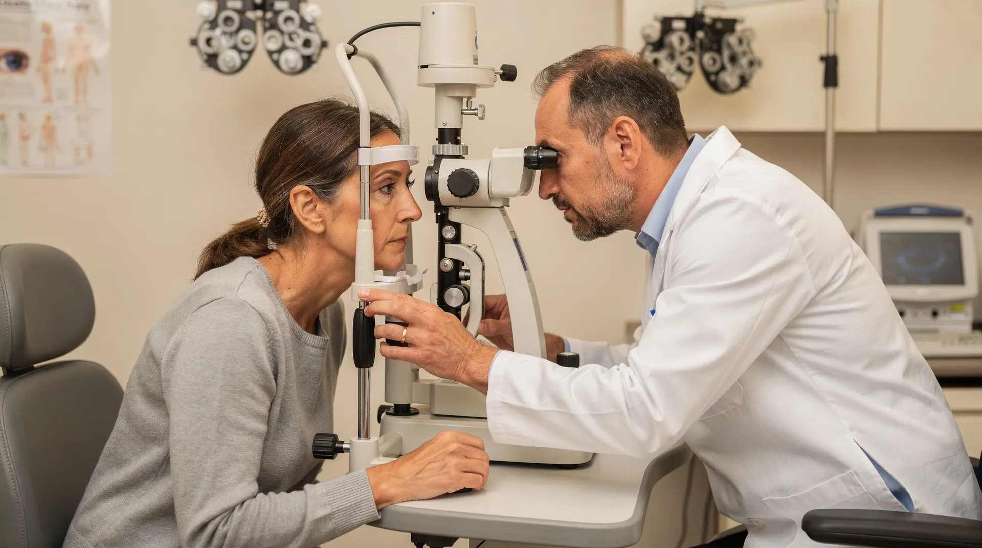
A glaucoma evaluation may include measuring eye pressure, examining the optic nerve, checking peripheral vision, assessing drainage angles, and reviewing other eye-health factors. The exact combination of tests depends on what the clinician needs to assess from your history and examination.\n\nCommon parts of the evaluation include:\n\n- Tonometry: Tonometry measures pressure inside the eye using an instrument called a tonometer.\n- Visual-field testing: A visual-field test measures peripheral vision and helps show whether peripheral vision has been lost. Testing therefore looks beyond whether your eyes feel comfortable at the moment. It checks pressure, structure, and visual function together so a qualified clinician can interpret the pattern rather than rely on one symptom or measurement.
An ophthalmic evaluation may also include a dilated eye exam. During a dilated eye exam, drops widen the pupils so an eye-care professional can use a special magnifying lens to examine the retina and optic nerve for signs of damage and other eye problems. Close-up vision may remain blurred for several hours after a dilated eye exam, so possible temporary near-vision blur is a practical consideration when planning the visit. The optic nerve is a bundle of more than 1 million nerve fibers connecting the retina to the brain, and a healthy optic nerve is necessary for good vision.
Pachymetry measures corneal thickness using an ultrasonic-wave instrument. A numbing drop is applied to the eye for pachymetry. Numbing drops may be applied to the eye for tonometry. Dilated eye examination, tonometry, pachymetry, and visual-field testing examine different aspects of eye health.
Up to one-half of patients with primary open-angle glaucoma are undiagnosed because diagnosing primary open-angle glaucoma often requires monitoring over years to document changes suggesting the disease. Primary open-angle glaucoma is the most common form of glaucoma in the United States. A single visit may be one part of a glaucoma assessment when findings need to be monitored over time.
Clinical findings guide glaucoma treatment categories
Glaucoma treatment aims to lower intraocular pressure and may include prescription eye drops, oral medicines, laser treatment, surgery, or a combination of approaches. Treatment is not selected from symptoms alone. Depending on the underlying cause of glaucoma, an eye doctor may recommend one or more treatments.\n\nThe broad categories include:\n\n- Prescription medicines: Eye drops and, in some circumstances, oral medicines may be used as part of a pressure-lowering plan.\n- Laser treatment: Trabeculoplasty is a laser treatment used for open-angle glaucoma, and other laser treatments can treat other types of glaucoma.\n- Surgery: Glaucoma surgery options include trabeculectomy, glaucoma implant surgery, and minimally invasive glaucoma surgery.\n- Combined care: More than one treatment category may be used when the underlying cause and clinical findings call for it.\n\nThese categories explain what may be discussed after an evaluation; they do not identify which option belongs in your situation. The clinician must interpret your eye pressure, optic-nerve appearance, visual-field results, drainage-angle findings, and other relevant information before recommending a plan. Do not start, stop, or change glaucoma medication or pursue a procedure based only on a symptom description.

Prepare the symptom and medical history for an evaluation
Your symptom and medical history can help an ophthalmic evaluation begin with the details that change how your concern is understood. Before arranging care, write down what you noticed and when it began. A brief record is enough.\n\nInclude these details:\n\n- Whether the change affects one eye or both eyes\n- Blurred vision, halos around lights, eye pain, headache, or eye redness\n- Whether symptoms began suddenly, appeared gradually, returned, or stayed present\n- Any change in side vision or sudden loss of vision\n- A family history of acute angle-closure crisis or other glaucoma\n- Topical or systemic medications you use\n- Any previous eye-pressure, optic-nerve, or visual-field findings you know about\n\nAn initial evaluation may include your history of blurred vision, halos, eye pain, headache, eye redness, family history of acute angle-closure crisis, and use of topical or systemic medications. Bring a medication list if you have one, and describe the timing in plain language rather than trying to assign a diagnosis.\n\nAdults over 60 should schedule annual eye exams. This general recommendation does not replace a prompt evaluation for a new or worsening symptom. We offer clinical and surgical glaucoma evaluations as part of our subspecialty eye care. Arrange an evaluation when your concern is gradual, recurring, unexplained, or associated with risk factors, unless sudden severe symptoms require emergency care instead.
Seek immediate help for sudden severe eye symptoms
Sudden severe eye symptoms require immediate qualified eye-care or emergency help. Seek emergency care right away for sudden, severe eye pain or pressure because acute angle-closure glaucoma can cause permanent vision loss in just a few hours.\n\nSeek immediate evaluation by an eye specialist for sudden vision loss, including partial or complete loss or sudden blurred or hazy vision. A sudden red eye, halos around lights, nausea, vomiting, or a severe headache occurring with eye symptoms can also be part of an acute angle-closure attack.\n\nUse this action sequence:\n\n1. Treat sudden onset as urgent. Do not wait to see whether every warning sign appears.\n2. Call an ophthalmologist or seek emergency care immediately. Sudden severe eye pain or sudden vision loss should not be handled as a routine appointment request.\n3. Go to the nearest emergency department if you cannot reach an eye doctor immediately. Do not wait for an online appointment.\n\nThis pathway applies to sudden or severe symptoms, not only to a confirmed glaucoma diagnosis. The purpose is to get immediate assessment when the pattern could threaten vision.

Use the symptom pattern to choose your next eye-care step
Your next eye-care step should match the symptom pattern. A gradual, recurring, unexplained change—or no obvious symptoms despite relevant risk factors—calls for an ophthalmic evaluation, because early open-angle glaucoma may be silent and symptoms cannot establish the diagnosis. Sudden severe pain, sudden vision loss, a red eye, halos, nausea, vomiting, or severe headache calls for immediate eye-care or emergency help instead.\n\nBring the timing, symptoms, family history, and medication information you recorded to the evaluation. A qualified eye-care professional must decide what the findings mean, whether glaucoma is present, and which treatment category—if any—fits your condition. Use this information to choose the urgency of care, not to diagnose yourself or select treatment.\n\nFor a non-emergency concern, take the next step toward an ophthalmic evaluation with DLV Vision.
Questions to ask about glaucoma symptoms and evaluation
Can glaucoma affect one eye or both?
Glaucoma can develop in one eye or both eyes. The pattern of symptoms does not establish whether glaucoma is present, so an ophthalmic examination is needed when you have unexplained or recurring vision changes or relevant risk factors.
Are halos around lights always an emergency?
Halos need immediate evaluation when they begin suddenly or occur with severe eye pain, a red eye, blurred or sudden vision loss, headache, nausea, or vomiting.
Is glaucoma curable?
There is no cure for glaucoma, and vision already lost from the disease cannot be restored. Treatment may help control the condition and often protect against serious additional vision loss; the appropriate approach depends on the examination findings and underlying cause.