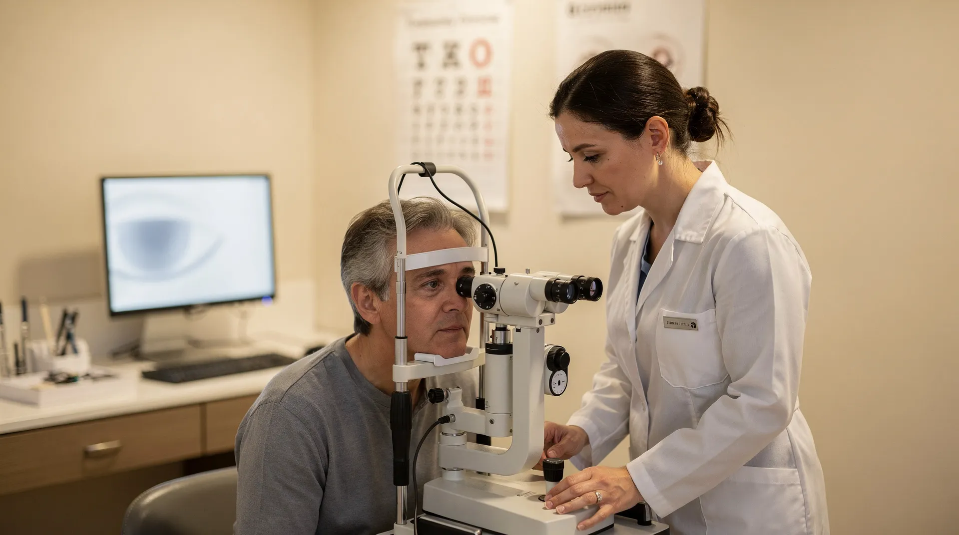
Glaucoma care begins with protecting the optic nerve
Glaucoma can damage the optic nerve and cause vision loss. The damage can reduce your vision permanently, often beginning with side vision before affecting what you see straight ahead. Without treatment, glaucoma can gradually reduce peripheral vision and may eventually cause blindness.
Eye pressure matters because fluid that does not drain normally can build up and damage the optic nerve. Increased pressure plays an important role in many cases, but glaucoma can also occur when eye pressure is normal or low. Researchers estimate that 50 to 80 percent of people with open-angle glaucoma have eye pressure higher than average, while others have normal or low pressure, and many people with high eye pressure never develop glaucoma (). A pressure reading therefore gives your ophthalmologist useful information without answering every question by itself.
The condition affects a large population. Open-angle glaucoma affects about 2.2 million Americans age 40 and over, while another two million may have it without knowing. A separate estimate places the total at more than 2.7 million people in the United States and more than 60 million worldwide. These figures use different populations and estimates, so they should not be added together.
Treatment is directed at controlling pressure and slowing further optic-nerve damage, not at restoring vision that has already been lost. In people at higher risk, pressure-lowering eye drops reduced the development of primary open-angle glaucoma by more than 50 percent; 4.4 percent of participants who received the drops developed glaucoma within five years, compared with 9.5 percent of participants who did not receive them. That result describes a higher-risk group and does not determine what treatment you need.
Your next decision should begin with the urgency of your symptoms and the need for objective testing. If you have no sudden severe symptoms, arrange an ophthalmology evaluation rather than trying to decide from a single pressure number or from how your vision feels. If sudden intense eye pain, blurred or red vision, nausea or vomiting, severe headache, or rainbow-colored halos appear, use immediate eye-care or emergency help instead.
A practical way to organize glaucoma care is to ask four questions:
- Could the symptoms represent an urgent angle-closure attack?
- What do pressure, visual-field, optic-nerve, drainage-angle, and imaging findings show?
- Which treatment category fits the documented disease status and pressure goal?
- What follow-up and cost questions need answers before you proceed?
The rest of this guide follows that order. It explains what the tests can show, how medicines, laser procedures, and surgery differ, why monitoring remains necessary, and which cost questions belong in your consultation.
A pressure reading is evidence, not the whole decision
Eye pressure is the force created by fluid inside your eye, and higher pressure increases the risk of glaucoma. A high reading does not, by itself, establish that you have glaucoma. Some optic nerves tolerate more pressure than others, and some people develop glaucoma even when their pressure is normal or low.
For most people, eye pressure above 21 is higher than normal. The number 21 is a general reference point, not a personal treatment target. The amount of pressure your optic nerve can tolerate differs from the amount another optic nerve can tolerate. Your ophthalmologist therefore interprets pressure alongside the optic nerve, visual field, drainage angle, cornea, medical history, and changes over time.
A single reading can also be affected by how the measurement is taken and by features of your eye. Corneal thickness, for example, can affect how pressure results are interpreted. A pressure result that looks reassuring does not rule out glaucoma, and a result above a general reference range does not prove that glaucoma has damaged your optic nerve.
Use the number as a reason to ask better questions, not as a diagnosis. Ask what the reading means for your optic nerve, whether the result should be repeated, which other tests are needed, and what findings would change the plan.
Normal-tension glaucoma is a form of open-angle glaucoma that can develop without increased eye pressure, and pressure-lowering treatments often help. That possibility is another reason an apparently normal pressure result cannot replace examination of the optic nerve and visual field.
If someone has told you that your eye pressure is high, arrange the evaluation that connects the pressure result with the rest of your eye findings. If your pressure is described as normal but you have concerning visual-field or optic-nerve findings, do not treat the number as permission to dismiss the concern. Let the complete examination determine the next discussion.
Silent changes and risk patterns can make evaluation worthwhile
Early open-angle glaucoma often has no warning signs, so you may not notice vision loss right away. Published U.S. estimates of how common the open-angle type is differ: one reports 9 in 10 people with glaucoma, while another reports that open-angle glaucoma affects about three times as many people as angle-closure glaucoma. The estimates may use different methods or definitions; the consistent point is that open-angle glaucoma is more common. Your vision may seem normal and your eye may not hurt even while changes are developing. As open-angle glaucoma progresses, it can affect side vision in one or both eyes.
Symptoms alone cannot separate glaucoma from other eye conditions or confirm how much damage has occurred.
A recognized risk factor or a family history of glaucoma can make an evaluation worthwhile even when your vision seems unchanged. Your risk history is one part of the appointment, not a diagnosis.
Before an appointment, write down information that can help your ophthalmologist interpret a quiet or gradual pattern:
- Your age and any family history of glaucoma.
- Whether you have noticed missing side vision, trouble navigating, or a gradual narrowing of what you see.
- Any previous pressure readings, visual-field tests, imaging, or eye examinations.
- Your current medicines and relevant medical conditions.
- Any past eye injury, eye surgery, or period when one eye changed differently from the other.
Do not wait for obvious vision loss before arranging routine evaluation if you have recognized risk factors or have been told that your pressure, optic nerve, or visual field needs monitoring. If sudden severe symptoms develop, follow the urgent pathway described later rather than waiting for a routine appointment.
What a glaucoma evaluation measures and why each test matters
A glaucoma evaluation combines your history with several views of how your eyes function and how their structures look. An initial examination may include eye-pressure measurement.
Each part answers a different question:
- History and vision checks: These establish your symptoms, previous findings, visual function, medicines, and relevant health information.
- Eye-pressure measurement: Tonometry measures the pressure inside your eye. The result is interpreted with the rest of your findings rather than used alone.
- Visual-field testing: This checks how well you detect objects away from the center of your vision without moving your eyes. It can show functional changes in side vision.
- Slit-lamp and fundus examination: These allow the eye-care professional to inspect the front and back of your eye and examine structures associated with glaucoma.
- Optic-nerve and retinal-nerve-fiber-layer assessment: These look for structural changes that may support a diagnosis or provide a baseline for monitoring.
Additional diagnostic testing may include central corneal-thickness measurement, visual-field evaluation, and imaging of the optic nerve, retinal nerve fiber layer, and macula. Corneal thickness gives context for interpreting pressure. Imaging records structural information, while visual-field testing records functional information. Comparing both types of information over time can make a change easier to identify than relying on symptoms alone.
Your appointment may not include every test at every visit. The ophthalmologist chooses the combination based on your history, examination findings, suspected type of glaucoma, previous results, and the need to establish or compare a baseline. Ask what each test is intended to clarify and which result would change your next step.
Practical planning can matter during testing. A dilated eye exam may leave vision blurry and sensitive to light for a few hours, so a person having the exam may need someone else to take them home. Ask the eye-care office whether dilation is planned and what arrangements are appropriate for that visit. The temporary effects of dilation are different from the permanent vision changes glaucoma can cause. Pressure measurement checks current eye pressure, visual-field testing checks visual function, and imaging or optic-nerve examination checks eye structure. Asking which baseline a repeat test is establishing can make the reason for testing clear even when symptoms have not changed.

How findings distinguish monitoring from treatment discussions
Some glaucoma suspects have no signs of damage. You may be considered a glaucoma suspect because of consistently high eye pressure, a suspicious optic-nerve appearance, thinning seen on imaging, or a suspicious visual-field result. Some glaucoma suspects have normal eye pressure.
The useful question is not simply whether you have been labeled a suspect. Ask which finding led to the label, whether the finding is present in one eye or both, whether it has changed, and what would move the plan from observation to treatment. A pressure reading, an optic-nerve appearance, an image, and a visual field each describe different parts of the risk picture.
Monitoring is active care. It gives your ophthalmologist repeated opportunities to check whether the findings that led to the label change over time. Structural changes in the optic disc and retina can be considered alongside functional changes measured through visual-field testing. The comparison is especially useful when your symptoms are absent or unchanged.
Risk and change are related but not identical. For a glaucoma suspect, the decision to treat depends on the pressure level and associated risk factors; if evidence suggests conversion to glaucoma, treatment may be considered. Medical therapy or laser trabeculoplasty can be discussed in some suspect situations, but the applicable choice remains tied to examination findings. When a visual-field defect is newly detected in a suspect, repeating the test can help determine whether the result persists. These are reasons to ask what the baseline result was and what later change would alter care.
The record can therefore separate three questions: what prompted concern, whether the finding is reproducible, and whether it is changing. A high reading may be risk information rather than proof of damage; conversely, normal pressure does not erase concern when structural or field findings are suspicious. A follow-up plan should identify which of those questions remains unanswered.
At the end of an evaluation, ask for a plain-language monitoring plan:
- Which finding is being watched?
- What is the baseline for pressure, visual field, optic nerve, or imaging?
- Which change would suggest progression?
- How often should you return?
- Which new symptom should prompt earlier contact?
Do not interpret monitoring as proof that nothing matters. It means the ophthalmologist is using repeated findings to decide whether the condition is stable, changing, or ready for a different discussion. Keep copies of your results when available so you can describe prior testing accurately if your care involves more than one office.
The treatment goal is pressure control and stable vision measures
Glaucoma treatment is intended to control eye pressure and slow further damage. It cannot restore vision already lost. The treatment goal is not a single pressure number that applies to everyone; it is a target range interpreted alongside the health of your optic nerve, retinal nerve fiber layer, and visual fields.
A glaucoma target pressure is an estimate that must be individualized and may be adjusted during the disease. The target can change when testing shows progression, when the level of optic-nerve damage changes the balance of risk, or when the current approach does not provide enough control or cannot be tolerated. Your ophthalmologist uses the pattern of findings to decide whether the target remains appropriate.
The AAO summary recommends an initial target pressure of at least 25 percent lower than pretreatment eye pressure when therapy is indicated. That 25 percent is a recommendation for an initial target in the guidance, not a personal instruction for you to calculate or pursue without an ophthalmologist. The appropriate target may be lower or otherwise adjusted according to your findings and disease course.
Use the treatment goal to structure your appointment. Ask:
- What pressure range is the plan targeting, and why?
- Which optic-nerve, retinal nerve fiber layer, or visual-field findings support that target?
- How will you know whether the target is being reached?
- What would count as progression?
- If the first approach is not enough or is difficult to tolerate, what alternatives could be discussed?
This approach keeps the conversation focused on measurable findings and the reason for each treatment category rather than on choosing a procedure in isolation.
Eye drops: pressure control that depends on continued use
Medical therapy is presently the most common initial intervention used to lower eye pressure. Eye-drop medications can lower pressure by decreasing how much fluid your eye makes or by helping your eye drain fluid more effectively. They work only while you are using them, so the treatment discussion includes both their expected pressure effect and whether you can use them as prescribed over time.
Effectiveness and side effects are considered together when an ophthalmologist chooses a regimen. The relevant questions include whether pressure reaches the target, whether the drops are tolerated, whether the schedule fits your daily routine, and whether testing shows stable or changing optic-nerve and visual-field findings. Your ophthalmologist may change the regimen when the response or tolerance changes.
Do not start, stop, combine, or change glaucoma medicine based on a general article. If you are already being treated, use the medicine as prescribed and tell your eye-care professional about missed doses, discomfort, burning, stinging, difficulty placing the drop, or any other problem that makes the plan hard to follow. A practical problem with the regimen is information your ophthalmologist can use when discussing alternatives.
Bring these questions to a medication discussion:
- What is the purpose of each drop?
- How does each drop affect fluid production or drainage?
- What schedule should you follow?
- What should you do if you miss a dose or cannot place the drop correctly?
- Which effects should be reported promptly?
- When will pressure and visual function be checked again?
The answer should connect the medicine to your documented pressure goal and follow-up plan. It should not be based only on whether your eyes feel different, because symptoms do not reliably show whether pressure control is working.
Laser treatment: another route to improve drainage
Laser treatment can help some types of glaucoma by improving fluid drainage. Laser trabeculoplasty is used for open-angle glaucoma. During the procedure, laser energy treats the drainage angle so it can work better over time and help lower eye pressure.
That makes follow-up part of the treatment decision rather than an optional extra. The ophthalmologist needs to see how your pressure responds and whether the rest of your glaucoma findings remain stable.
Laser trabeculoplasty may take about 6 to 8 weeks to fully lower eye pressure. If your pressure is still too high three months after the procedure, more treatment may be needed. Those time points describe when the response may be assessed; they do not prescribe a personal appointment schedule.
A 2024 report described a comparison in which glaucoma progressed 29 percent slower in patients treated with laser than in patients treated with eye drops. In the reported six-year follow-up analysis, the estimated proportion of eyes with fast progression was 14 percent in the laser group and 25 percent in the eye-drop group. Those figures describe the reported comparison and should not be converted into a prediction for your eyes.
Ask whether your glaucoma type, drainage angle, pressure pattern, optic-nerve findings, and ability to use eye drops make laser treatment a reasonable topic for discussion. Also ask how the response will be measured, what happens if pressure remains above target, and whether you would still need medicine or another procedure.
Surgery: improving outflow when other approaches are not enough
When medicines and laser treatment are not enough to control eye pressure, an ophthalmologist may discuss glaucoma surgery. Traditional procedures such as trabeculectomy or a tube shunt implant may create a new drainage channel for fluid to leave the eye. Incisional surgery can also improve fluid outflow through a small surgical opening in the eye’s drainage system or sclera, the white part of the eye.
Some patients may be offered minimally invasive glaucoma surgery. The category is broader than a single procedure, so the relevant questions concern the specific operation being proposed, its purpose, the pressure target, the expected monitoring, and what other treatment may still be needed.
Surgery is intended to help control pressure and protect remaining vision; it does not cure glaucoma or undo vision loss that has already occurred. The need for surgery is determined by your findings and response to other treatment, not by the presence of a glaucoma label alone.
If surgery enters the conversation, ask for clear answers to these questions:
- What pressure problem is the procedure intended to address?
- Why is this operation being considered now?
- What other treatment categories remain available?
- What type of drainage route or opening would the procedure create?
- What follow-up testing will show whether it is working?
- Could you still need drops, laser treatment, or another procedure?
- Which symptoms or changes after treatment require prompt contact?
A surgical consultation should leave you with a named procedure category, a reason it fits the documented findings, and a monitoring plan. If those points are unclear, ask the ophthalmologist to restate them in everyday language before making a decision.
How clinicians match the treatment path to the findings
Because progression can vary, an ophthalmologist individualizes treatment and follows it closely. The pressure target may change over time. That means the treatment path is a sequence of decisions connected to testing, response, and follow-up rather than a permanent ranking of medicines, laser, or surgery.
| Treatment category | Main purpose | Questions that shape the discussion |
|---|---|---|
| Eye drops | Lower pressure by decreasing fluid production or helping fluid drain better | Is the pressure reaching target? Can you use the drops consistently? Are effectiveness and tolerance acceptable? |
| Laser trabeculoplasty | Improve drainage for some types of glaucoma, especially open-angle glaucoma | Is the drainage angle suitable? How will pressure response be checked? Could you still need drops or additional treatment? |
| Glaucoma surgery | Create or improve a drainage route when medicines or laser treatment are not enough | What procedure is being proposed, why now, what pressure goal applies, and what follow-up will be required? |
The table is a discussion framework, not a recommendation for your care. An ophthalmologist uses your disease progression, pressure, optic-nerve damage, visual-field results, tolerance, and other risk factors to determine which category should be discussed.
Every category has a boundary. Drops depend on continued use. Laser treatment may not lower pressure enough and may require additional treatment. Surgery may be considered when less invasive approaches do not provide enough control, but it cannot restore lost vision. Ask your ophthalmologist to compare the alternatives using your own findings, pressure target, expected monitoring, and tolerance of the proposed plan.
What glaucoma treatment may cost and what changes the price
Glaucoma treatment costs vary by procedure, facility, anesthesia, operating-room charges, medicines, insurance or Medicare coverage, and whether you need additional treatment. One U.S. estimate lists laser glaucoma procedures at between $1,000 and $2,000, more involved surgeries at around $5,000, traditional trabeculectomy at about $4,200, and microshunt procedures closer to $6,300. The estimate places anesthesia and operating-room fees in addition to those amounts.
These figures are published estimates, not personalized quotes. They describe different procedure categories and should not be used to predict your bill. A laser procedure and an incisional operation may involve different facility arrangements, professional fees, follow-up needs, and coverage rules. Ask for the total expected charge and the part you may owe before scheduling nonurgent care.
Coverage and longer-term treatment can change the financial picture. Ask your insurer or Medicare what applies to your diagnosis, procedure, facility, anesthesia, medicines, and follow-up rather than assuming that every charge is handled the same way.
A claims-data analysis found that glaucoma-specific pharmacy costs were lower after laser trabeculoplasty than after additional medications during the 24 months after the procedure, while overall glaucoma-specific treatment costs were comparable. The analysis did not account for individual clinical findings or distinguish among different laser treatments and application patterns. Treat it as a comparison of claims patterns, not as a promise that laser treatment will cost less for you.
Patients can make published glaucoma cost estimates easier to compare by recording each expense as a separate line item: the professional fee, facility charge, anesthesia or operating-room charge, medicines, diagnostic testing, follow-up, and possible additional treatment. Each line item can be marked as included, billed separately, or unknown. A worksheet using those named categories can show whether a quoted amount covers more than one procedure charge or covers only the procedure itself. Separate one-time charges from recurring expenses when making the worksheet. Medicines and follow-up can continue beyond the initial procedure, while a procedure estimate may not include later care. Ask the billing office which amounts are known before treatment and which amounts depend on later testing or additional treatment. Coverage questions require plan-specific answers. Ask the insurer or Medicare whether coverage and applicable authorization rules differ for the named procedure, facility, medicines, diagnostic testing, or follow-up. A published estimate cannot determine which treatment is clinically appropriate or predict an individual patient’s final responsibility. Use the estimate to prepare a focused financial discussion after an ophthalmologist explains the treatment options that fit the documented findings.
Use these questions to make a cost conversation specific:
- What procedure or treatment category is being considered?
- Which professional, facility, anesthesia, medication, imaging, and follow-up charges are separate?
- What is the estimated total before insurance or Medicare?
- What amount may remain after coverage, and could prior treatment be required?
- Would additional laser treatment, medicines, or surgery create later charges?
- Which costs are expected only if testing shows that more treatment is needed?
Request written estimates when available, but keep the clinical decision tied to your findings and treatment goal rather than choosing an option by price alone.
Follow-up is how treatment response becomes visible
You generally cannot tell by symptoms whether glaucoma treatment is working. You may not feel eye pressure rising or notice early vision loss, and side effects such as burning or stinging do not show whether pressure has reached its target. Follow-up testing is therefore used to check pressure, side vision, the optic nerve, retinal nerve fiber layer, and other findings that help show whether the condition is stable or changing.
Follow-up visits are generally scheduled two to four times per year, although your ophthalmologist may want to see you more or less often. An AAO consensus table gives approximate follow-up intervals ranging from 1–2 months to 12 months, depending on pressure control, damage progression, and how long control has been maintained. The interval belongs to your clinical situation; do not turn either range into a personal schedule without an ophthalmologist. Many people with glaucoma need additional treatment over time, and monitoring may be lifelong. A later treatment discussion can reflect changing findings or the need for additional pressure control.
Bring a written question list to the consultation:
- What is the working diagnosis, and which findings support it?
- What is the treatment goal for pressure, optic-nerve structure, and visual-field function?
- What alternatives are available, and how do their benefits and risks differ for your findings?
- How will you know whether the chosen treatment is working?
- Which tests will be repeated, and what change would indicate progression?
- When should your next appointment occur?
- What symptoms or changes should prompt contact before that appointment?
- What should you do if a drop is difficult to use or a treatment is not tolerated?
You should receive information about the disease, treatment goals, your condition, and the relative benefits and risks of alternatives so you can participate in planning care. Leave with the next test, the next appointment, and the reason for each.

When sudden symptoms require immediate eye care
A suddenly blocked drainage angle can cause an acute angle-closure attack, which is a true eye emergency. Seek immediate eye-care or emergency help for sudden intense eye pain, blurred vision, red eyes, or nausea. Severe eye or brow pain, severe headache, vomiting, and rainbow-colored halos around lights are additional symptoms reported with angle-closure glaucoma.
Angle-closure glaucoma may have no symptoms before an attack. Early attack symptoms can include blurred vision, halos, mild headaches, or eye pain, so contact an ophthalmologist as soon as possible if these symptoms appear. Do not wait for a routine appointment to see whether severe or rapidly developing symptoms pass.
If you are unsure whether a sudden eye problem is urgent, contact a qualified eye-care professional or the responsible emergency authority promptly. The practical boundary is speed: sudden severe symptoms need immediate assessment, not a diet change, exercise plan, online self-diagnosis, or a decision based on an old pressure reading.
If you are already being treated for glaucoma, take your medicine every day and see your eye-care professional regularly. Tell the office if you cannot follow the schedule or if the medicine causes a problem so the treatment team can address it.
No special diet or exercise regimen has been shown to prevent glaucoma. A healthy lifestyle can help you manage diabetes and high blood pressure, which can increase glaucoma risk, but lifestyle measures do not replace pressure checks, visual-field testing, optic-nerve evaluation, or prescribed treatment. Use them as general health measures, not as a substitute for glaucoma care.
Arrange the next glaucoma conversation
Your next step depends first on urgency. Sudden severe eye symptoms call for immediate eye-care or emergency help. Without that warning pattern, arrange an ophthalmology evaluation that connects your symptoms and risk history with pressure, visual-field, optic-nerve, drainage-angle, corneal-thickness, and imaging findings.
Bring your medication list, previous test results, family history, and a short description of any vision changes. Ask which treatment goal applies to you, why a medicine, laser procedure, surgery, or monitoring plan is being discussed, how response will be measured, how often follow-up is expected, and which charges may affect your share of the cost. A qualified ophthalmologist must determine your individual diagnosis, suitability, and treatment plan.
We offer clinical and surgical glaucoma evaluations. Use the evaluation conversation to turn uncertainty into a documented next step rather than trying to choose treatment from symptoms or cost estimates alone.
If you have sudden severe symptoms, seek immediate qualified help instead of waiting for a routine contact process. If your concern is nonurgent, prepare your questions and arrange the appropriate ophthalmology conversation.
Questions patients commonly ask before glaucoma care
Can you have glaucoma with normal eye pressure?
Yes. Glaucoma can occur when eye pressure is normal or low, and a high pressure reading does not by itself mean that glaucoma is present. An ophthalmologist interprets pressure alongside the optic nerve, visual field, drainage angle, cornea, imaging, and changes over time.
What should be included in a glaucoma treatment cost estimate?
Ask whether the estimate includes the professional fee, facility, anesthesia, operating-room charges, medicines, diagnostic testing, and follow-up. Published estimates list laser procedures at $1,000 to $2,000, more involved surgeries around $5,000, traditional trabeculectomy about $4,200, and microshunt procedures closer to $6,300, before anesthesia and operating-room fees. Your insurance or Medicare coverage and any need for additional treatment can change what you owe.
Can glaucoma treatment restore vision that has already been lost?
No. Medicines, laser procedures, and surgery are intended to control eye pressure and slow further optic-nerve damage; they cannot restore vision already lost. Early evaluation, prescribed treatment, and ongoing monitoring can help protect remaining vision.
Why are follow-up tests needed if my eyes feel normal?
Glaucoma and rising eye pressure often cause no noticeable symptoms early, so how your eyes feel cannot reliably show whether treatment is working. Follow-up testing checks pressure, side vision, the optic nerve, retinal nerve fiber layer, and other findings so an ophthalmologist can identify stability or progression and adjust the plan when needed.