Recognize the growth that can reach the cornea
An eye pterygium is a raised, blood-vessel-containing growth that begins in the conjunctiva, the thin membrane on the eye’s surface, and extends onto the cornea. It may affect one eye or both eyes. The growth is often fleshy and triangular, and it may range from a fine, lightly elevated early lesion to a thicker, more vascular growth.
The cornea is the clear front surface of the eye. A pterygium matters because it can extend across that surface rather than remaining only on the white part of the eye. A small growth may cause no symptoms, while a larger or advancing growth may irritate the eye or affect vision if it reaches the visual axis.
An eye pterygium can look noticeable, but appearance alone cannot tell you how far it extends or whether another condition is responsible. Use the visible growth as a reason to arrange an eye evaluation, not as a diagnosis. The practical first question is whether the growth is confined to the surface or has begun affecting the cornea, refraction, or vision. A pterygium may occur on the nasal or temporal side of both eyes or at only one location, so a one-sided appearance does not rule out a pterygium.
Separate a pterygium from a pinguecula
A pterygium is a fleshy growth that can extend onto the cornea. Both are surface-eye growths, so they can look similar at first glance, but their relationship to the cornea is the practical distinction.
A pinguecula remains confined to the conjunctiva in the description used for comparison. A pterygium can cross onto the cornea and take on a wedge-like or wing-shaped appearance. A pterygium may start as a pinguecula, but a visible bump is not enough to establish that this has happened.
You may notice a yellow, white, pink, or fleshy area near the inner corner of the eye. Color and shape can provide useful observations to share during an examination, but they do not identify the growth by themselves. Arrange qualified eye care if the area is new, changing, irritated, or associated with a change in vision.
Who is more likely to develop pterygium
Pterygium risk is associated with substantial outdoor exposure and with sunny, dusty, sandy, windy, or dry environments. Sunlight, including ultraviolet light, wind, and dust are believed to contribute to both pinguecula and pterygium. The cause is not fully settled; inflammation, tear-film abnormalities, immune abnormalities, and a possible viral stimulus have also been proposed.
Pterygium is more common near the equator and in older adults, and risk increases with age. Pterygium is rare in children. An estimate suggests that about 12% of Australian men over 60 years have pterygium; this figure applies only to that population and does not describe prevalence among U.S. adults.
Your outdoor habits do not determine whether a growth is present, but they can help explain why an eye professional may ask about sun, wind, dust, and dryness. Protection is a practical step while you arrange or attend an evaluation:
- Wear wraparound sunglasses in bright outdoor conditions.
- Use wraparound glasses, goggles, or other protective eyewear in dry or dusty settings.
- A wide-brimmed hat can add protection from sunlight.
Sunglasses that block 100% of UVA and UVB rays or a wide-brimmed hat were reported as measures that can significantly reduce pterygium risk. These measures protect against associated exposure; they do not identify or remove an existing growth.

How pterygium symptoms and vision changes can develop
Pterygium symptoms may be absent at first, or they may begin as surface irritation. Early pterygia are usually asymptomatic, but a growth can disturb wetting of the eye’s surface and cause burning, itching, or tearing. An inflamed pterygium may also cause redness or irritation.
Symptoms that deserve attention include:
- Persistent burning, itching, tearing, or redness.
- A foreign-body sensation that does not settle.
- A visible wedge-shaped membrane that appears to be advancing toward the cornea.
- Blurry or distorted vision.
- A change that seems related to the growth rather than to a brief environmental trigger.
Vision may be affected when a pterygium reaches the cornea or visual axis. Corneal changes can include regular or irregular astigmatism, which may make vision blurry or distorted. A pterygium larger than 3 mm may induce astigmatism. Lesions larger than 3.5 mm are likely to be associated with more than 1 diopter of astigmatism and often blur uncorrected vision, although the literature is unclear about how effectively surgery corrects that astigmatism.
A pterygium may look like a wedge-shaped, translucent membrane ranging from white to pink. A Stocker line may occur at its leading edge on the cornea. Those descriptions can help you explain what you see, but they cannot replace an examination.
Among surgically excised pterygia, reported concurrent ocular surface squamous neoplasia ranged from 1.7% in South Florida to as high as 9.8% in Australia. That range applies to reported surgically excised cases in those locations; it is not a general percentage for every visible eye growth. A changing or unusual growth should therefore be assessed rather than judged from appearance alone. Other documented features of a pterygium can include vascular straightening in the direction of the advancing head and degenerative changes such as cystic changes. A complete eye examination is needed to assess vascular straightening, cystic changes, and visual or refractive effects.

How pterygium may remain or change
A pterygium may stay small, or it may grow over part of the cornea and affect vision. The pace and extent of change are not something you can reliably determine by looking at the eye from day to day.
A pterygium does not go away on its own. Lubricating care may ease irritation, and protective eyewear may reduce exposure associated with the condition, but those measures do not remove the tissue. Eye surgery is the way to get rid of the growth.
For practical monitoring, note when you first noticed the area and whether its appearance, irritation, tearing, or vision has changed. Arrange an examination for a suspected pterygium even if symptoms are mild, particularly when the growth is changing. A qualified eye professional can determine whether observation remains reasonable or whether corneal or visual effects require a different discussion. A pterygium can remain visible while irritation varies, so temporary symptom relief does not establish that a pterygium has disappeared. Lubricating care may ease irritation, but lubricating care does not remove a pterygium. A new or persistent change in clarity, distortion, or eye comfort can warrant examination even when the visible area seems unchanged. A dated record of when the growth was noticed and when symptoms or vision changed gives an eye professional a fuller history than a single observation. The duration of a visible growth cannot confirm its identity or show how far it extends over the cornea.
What an eye examination can clarify
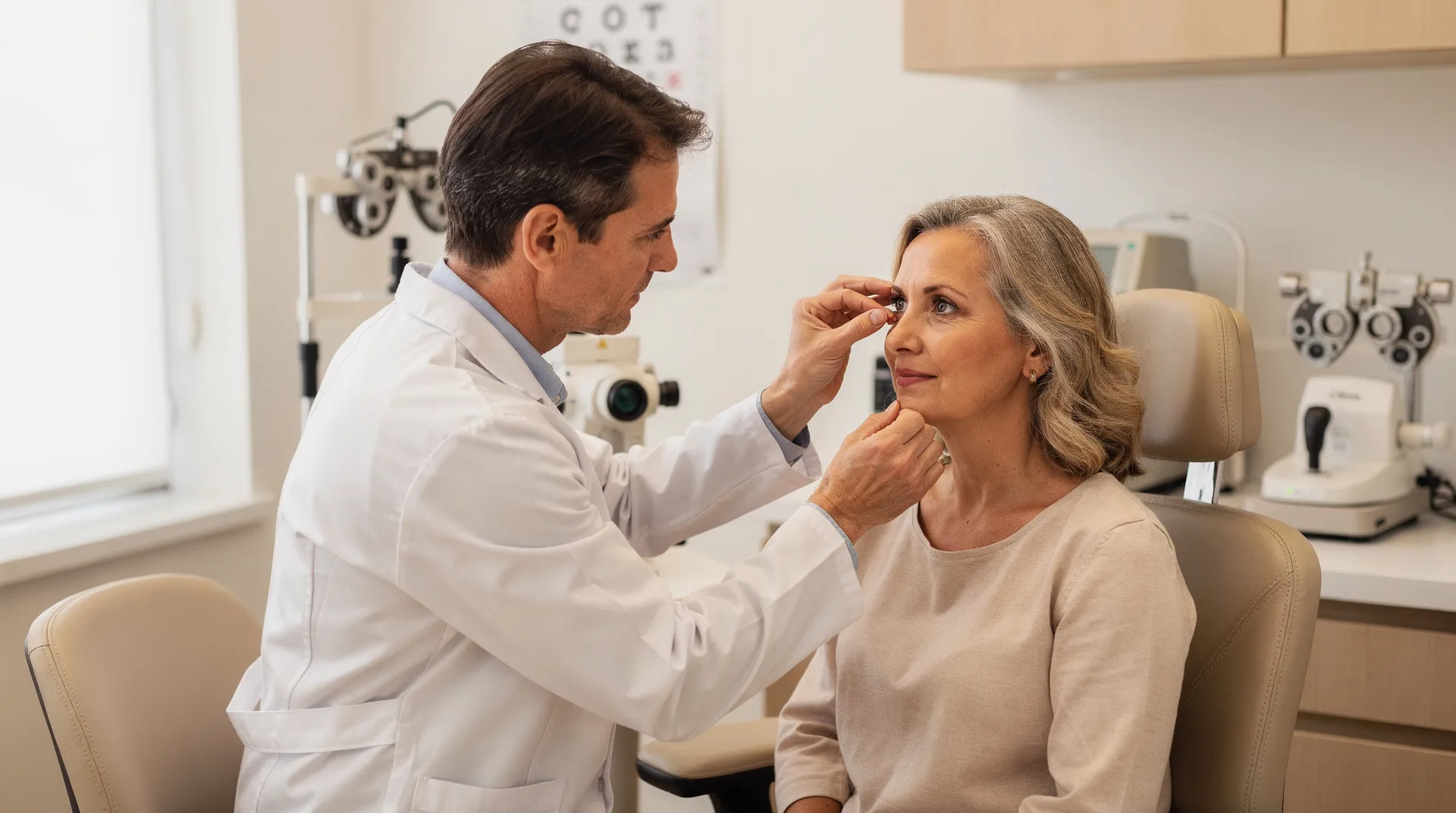
A pterygium examination can establish more than a photograph or mirror check. Diagnosing pterygium requires an eye-care professional’s examination, including slit-lamp inspection of the growth and the area where it may extend onto the cornea.
A complete eye examination can assess:
- Whether the growth is consistent with a pterygium or needs consideration of another diagnosis.
- How far the tissue extends across the cornea.
- Whether the surface is inflamed or irregular.
- Whether vision or refraction has changed.
- Whether the growth is affecting the visual axis.
Visual acuity, manifest refraction, and corneal topography can help determine a pterygium’s visual impact. The examination may therefore connect what you feel—such as irritation or blur—with what is happening on the eye’s surface.
Do not assume that an eye growth is a pterygium from appearance alone. Arrange routine qualified eye care for a stable suspected growth, and move to prompt care when vision changes, pain, marked redness, injury, or rapid worsening is present. After a pterygium is excised, histopathologic confirmation is routine and can help rule out malignancy. Histopathologic confirmation applies to removed tissue; histopathologic confirmation is not a home test or a substitute for the initial eye examination. An eye examination therefore answers two separate questions: whether a visible growth is consistent with pterygium and whether the growth is affecting the cornea, refraction, or vision enough to inform a treatment discussion.
Protect the eye and ease symptoms while monitoring
Pterygium symptom care focuses on limiting associated exposure and easing irritation; it does not remove the growth. Early-stage pterygia are often managed conservatively with lubricating medication and protective eyewear.
You can use a simple protection-and-observation routine:
- Use wraparound sunglasses in the sun.
- Use wraparound glasses, goggles, or other protective eyewear in dry, dusty conditions.
- Use artificial tears as directed on the product label when dryness or irritation is part of the pattern.
- Record whether burning, tearing, redness, or the foreign-body sensation improves, persists, or worsens.
- Arrange an eye examination rather than using symptom relief as proof of a diagnosis.
Artificial tears can lubricate the eye in dry conditions. Lubricating drops or ointments may ease minor irritation. For severe inflammation, a specialist may prescribe a short course of steroid eye drops; those medications ease symptoms and are not a cure.
Do not assume that comfort after lubrication means the growth has stopped or disappeared. If symptoms continue, recur, or occur with a change in vision, take that pattern to qualified eye care. That distinction matters when symptoms change. A temporary improvement after drops should therefore be reported as part of the pattern rather than treated as proof that the growth has stopped. Short courses of topical corticosteroids can reduce inflammation, but long-term use is not recommended.

When pterygium removal may be considered
Pterygium removal may be considered when the growth causes significant irritation, affects vision, grows aggressively, or creates another documented problem. Surgery is not an automatic response to every visible growth; the decision depends on what an examination shows and how the condition affects you.
Reasons an ophthalmologist may discuss removal include:
- Persistent or serious eye irritation.
- Reduced vision from astigmatism or extension toward the visual axis.
- Growth that is affecting the cornea.
- Difficulty wearing contact lenses.
- Symptomatic cystic changes.
- Restricted eye movement.
- A cosmetic concern after the clinical situation has been assessed.
A pterygium can grow back after removal. One reported recurrence estimate was between 30% and 40%, with recurrence more likely among people under 40 years; that figure is one reported estimate, not a universal result for every operation. Another report stated that recurrence decreased to 5%–10% with conjunctival flap or graft supplementation. Without an autograft or amniotic membrane transplant, recurrence was reported as high as 50% within 4 months and 97% within 12 months.
Those figures should not be combined into one personal forecast. Technique, graft use, follow-up period, patient and lesion characteristics, and the definition of recurrence can change the result. Ask the examining ophthalmologist to explain which factors apply to the proposed procedure.
What surgery and recurrence evidence can—and cannot—show
Pterygium surgery removes the growth, but recurrence remains a major challenge. Simple excision that leaves bare sclera has been reported to have recurrence risk upwards of 80%, while tissue grafting has a lower reported recurrence risk. A conjunctival autograft uses tissue from another part of the eye to cover the excision site; an amniotic membrane graft uses donor amniotic membrane for the remaining exposed area.
Reported recurrence figures vary by technique and study. Bare-sclera excision has ranged from 24% to 89%, while conjunctival autograft has ranged from 2% to 40% in several prospective studies. In a Cochrane review, recurrence at 3 months ranged from 0% to 16.7% after conjunctival autograft and from 4.76% to 26.9% after amniotic membrane transplant. At 6 months in that review, recurrence ranged from 3.33% to 16.7% after conjunctival autograft and from 2.6% to 42.3% after amniotic membrane transplant; conjunctival autograft had a 46% lower recurrence risk, but uncertainty remained about adverse events and meaningful visual-acuity changes.
These are not interchangeable percentages. The first ranges describe different techniques and reports; the Cochrane figures describe recurrence in treatment groups at specified follow-up points. A broader estimate of 30%–40% after removal and the reports of recurrence as high as 50% within 4 months or 97% within 12 months without an autograft or amniotic membrane describe other conditions and study contexts. No technique is universally accepted because recurrence rates vary.
Reported complications include corneal scarring, corneal perforation, strabismus, a nonhealing defect, scleral melt, and scleral dellen. Use the examination findings, the growth’s effect on vision, the proposed graft or technique, and the expected follow-up plan to make the surgical discussion specific to your situation.
What reported pterygium surgery costs mean
Pterygium surgery cost varies by treatment setting in that report. A report published on May 26, 2025, gave these average amounts:
- $1,795 for office-based pterygium surgery.
- $3,813 at a local surgical facility.
- $5,562 at a metropolitan-area hospital.
The figures are reported averages for different treatment settings, not a patient-specific price. They show that the setting was associated with different average costs ; they do not tell you what your own procedure, evaluation, anesthesia, facility services, graft, or follow-up will cost.
The same report described outcomes for office-based surgery in its own group: at 1 year, granuloma occurred in 3.8% of eyes, dellen in 3.1%, recurrence in 2.0%, and ptosis in 2.6%; no infections were reported. At 6 months, 94% of patients reported cosmetic satisfaction.
For a useful cost conversation, ask which setting is being considered and which services are included in the quoted amount. A practice can provide the terms that apply to your proposed care; the published averages above cannot substitute for an individualized estimate. The report does not provide a patient-specific quote or establish what every facility includes. Before relying on a figure, clarify whether the estimate covers the procedure, anesthesia, facility services, any graft, and follow-up visits. The answer may depend on the proposed surgical plan and the examination findings, so keep these published amounts separate from the estimate for the care being discussed.

Seek prompt qualified care for warning signs
Warning signs around a suspected pterygium require prompt qualified eye care rather than routine observation. Concerning eye symptoms include intense pain, a change in vision, blurry vision, very red or watery eyes, an object stuck in the eye, or serious eye injury or trauma.
Seek prompt evaluation if you notice any of the following:
- Sudden or significant change in vision.
- Intense pain.
- Marked redness or watering.
- An object stuck in the eye.
- A serious injury, including a hard blow to the eye.
- Rapidly worsening symptoms or a growth that seems to change quickly.
These signs do not prove that a pterygium is responsible. They indicate that the eye needs timely assessment. Do not delay care while trying to compare the growth with photographs or decide whether it looks like a pinguecula. If an injury or object in the eye is involved, avoid trying to diagnose the surface growth yourself and seek qualified help promptly.
Use your symptoms and changes to begin the next eye-care conversation
Your next eye-care step should follow the pattern you can describe, not the appearance of the growth alone. A mild, stable concern without warning signs can lead to a routine comprehensive eye evaluation, while persistent irritation, a changing growth, or vision changes should move that evaluation forward. Sudden vision changes, intense pain, marked redness or watering, an object in the eye, serious injury, or rapidly worsening symptoms call for prompt qualified care.
Before you begin the conversation, write down:
- When you first noticed the growth or surface change.
- Whether it is in one eye or both.
- Whether its size, color, irritation, tearing, or redness has changed.
- Whether vision is blurry, distorted, or newly different.
- Your outdoor, sun, wind, dust, or dry-environment exposure.
- Any contact-lens difficulty or prior eye injury.
An examination can determine whether the growth is a pterygium, assess its corneal and visual effects, and explain whether observation, symptom-focused care, or removal belongs in the discussion. A qualified professional must decide whether a particular evaluation or treatment is suitable for your eyes.
Questions patients commonly ask about eye pterygium
Can an eye pterygium go away without surgery?
No. A pterygium does not go away on its own, although artificial tears and protective eyewear may ease irritation or reduce exposure-related aggravation. Surgery is the treatment that removes the growth, and an eye-care professional must determine whether removal is appropriate.
Is a pterygium an emergency?
A stable, mild surface growth without warning signs may be evaluated through routine eye care. Seek prompt qualified eye care for sudden or significant vision changes, intense pain, marked redness or watering, an object in the eye, serious injury, or rapidly worsening symptoms. These signs do not prove that a pterygium is responsible, so avoid relying on appearance alone.
What should I ask about the cost of pterygium surgery?
Ask which treatment setting is planned and whether the estimate includes the evaluation, procedure, anesthesia, facility services, any graft, and follow-up. A report published on May 26, 2025, found average costs of $1,795 for office-based surgery, $3,813 at a local surgical facility, and $5,562 at a metropolitan-area hospital; these were setting-specific averages, not a patient-specific price.
Can a pterygium come back after it is removed?
Yes. Recurrence estimates vary substantially by surgical technique, graft use, follow-up period, and the patients and lesions studied. Reported figures have ranged from 0% to 16.7% at three months after conjunctival autograft in one review, where the denominator was the treated conjunctival-autograft groups, and from 4.76% to 26.9% after amniotic membrane transplant in that review’s treated groups. Those percentages are not a personal forecast; ask the ophthalmologist to explain the recurrence evidence for the proposed procedure.