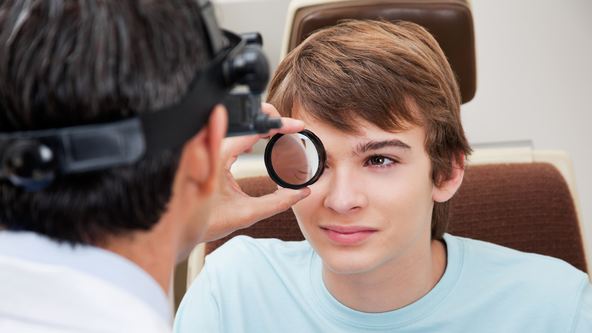
Optomap is a digital retinal imaging test that scans up to 82% of the retina in seconds without eye drops. Dilation uses eye drops to widen the pupil so an eye doctor can view the retina directly through a lens. Both methods check for the same conditions, including diabetic retinopathy, macular degeneration, and retinal tears. Optomap works faster and skips the blurry after-effects of drops, but some patients still need traditional dilation for a complete view. At DLV Vision, the clinical team uses Optomap imaging as the primary retinal screening tool for most patients across Ventura County and Los Angeles County.
Quick Takeaways
- Optomap is a wide-field digital photo of the retina taken without dilating drops, usually completed in under 30 seconds.
- Dilation uses eye drops to enlarge the pupil, giving the doctor a direct, hands-on view of the retina and its edges.
- Urgency: Neither test is an emergency procedure, but skipping annual retinal screening delays detection of silent eye diseases.
- Treatment approach: Optomap and dilation are screening tools, not treatments — findings guide next steps like monitoring, laser therapy, or referral to a specialist.
- When to schedule: Adults should have a retinal screening at least once a year, or sooner if they have diabetes, high blood pressure, or a family history of eye disease.
What Is Optomap Retinal Imaging?
Optomap is a widefield digital scanning laser device that captures a single image of most of the retina, the light-sensitive tissue lining the back of the eye. The scan takes less than a second per eye and does not require dilating drops for the majority of patients. The image displays instantly on a screen, so the doctor can review it with you during the same visit.
How Does Optomap Compare to a Standard Photo?
Traditional retinal cameras capture a narrow, central section of the retina. Optomap captures a much wider field, roughly 200 degrees, which includes the peripheral retina where tears and early diabetic changes often start. A wider view means fewer areas go unchecked.
What Is Pupil Dilation?
Dilation is the traditional method eye doctors use to examine the retina. The doctor places drops in each eye that relax the iris muscle and widen the pupil. Once the pupil is fully open, the doctor uses a bright light and handheld lens to look directly into the eye.
What Does Dilation Feel Like Afterward?
Dilated pupils let in more light and struggle to focus up close for several hours. Most patients experience light sensitivity and blurry near vision for four to six hours after the drops are administered. Many patients need sunglasses and cannot safely drive themselves home during this window.
Why Do Eye Doctors Recommend Retinal Screening?
Retinal disease often develops silently. Diabetes damages the small blood vessels in the retina, a condition called diabetic retinopathy, and this damage can progress for years without symptoms. High blood pressure, high myopia (severe nearsightedness), and a family history of retinal detachment also raise the risk of retinal changes. Left untreated, these conditions can cause permanent vision loss, including blind spots, distorted vision, or complete retinal detachment.
A comprehensive eye exam, using either Optomap imaging or dilation, is the only way to detect these changes early. This is why a comprehensive eye exam remains the gold standard recommended by ophthalmologists and optometrists alike. Early detection allows treatments such as laser therapy, injections, or monitoring to begin before vision is permanently affected.
Who Is at Higher Risk for Retinal Problems?
Patients with diabetes, uncontrolled blood pressure, or a family history of macular degeneration face the highest risk. Adults over age 60 and patients with high nearsightedness also need more frequent retinal screening, according to the American Academy of Ophthalmology.
What Symptoms Mean You Need a Retinal Exam Sooner?
Most retinal disease has no early symptoms, which is exactly why screening matters. When symptoms do appear, they include new floaters, flashes of light, a shadow or curtain across your vision, or sudden blurriness in one eye.
See an eye doctor immediately if you notice sudden vision loss, a sudden increase in floaters, flashes of light, or a dark curtain moving across your field of vision. These signs can indicate a retinal tear or detachment, which requires urgent evaluation.
What Can Optomap See That Dilation Cannot?
Optomap and dilation both examine the retina, but they differ in coverage, comfort, and speed. The table below compares the two methods side by side.
| Feature | Optomap Imaging | Traditional Dilation |
| Retinal coverage | Up to 82% of the retina in one image | Central and mid-peripheral retina, viewed directly |
| Eye drops required | No, for most patients | Yes, dilating drops widen the pupil |
| Time required | Under 30 seconds per eye | 20–30 minutes for drops to take effect, plus exam time |
| Blurry vision after | None | 4–6 hours of light sensitivity and near blur |
| Best for | Routine annual screening, quick documentation | Detailed exam of the extreme periphery, some retinal conditions |
Can Optomap Replace Dilation Completely?
Optomap replaces dilation for most routine annual screenings. However, some patients with diabetes, prior retinal detachment, or unclear Optomap results still need traditional dilation so the doctor can view the extreme edges of the retina directly. The National Eye Institute notes that a dilated exam remains an important diagnostic tool for detecting diabetic eye disease in certain cases.
Optomap or Dilation: Which Method Should You Choose?
Choose Optomap if you want a fast, comfortable screening with no drops and no driving restrictions afterward. Choose dilation, or accept your doctor’s recommendation for dilation, if you have diabetes, a personal or family history of retinal detachment, or if your Optomap image shows an area that needs a closer, direct look.
Do Eye Doctors Prefer Dilation or Optomap?
Most eye doctors now prefer Optomap as the first-line screening tool because it covers more retina, saves time, and improves patient comfort. Dilation remains the preferred method when a doctor needs to examine a specific area in greater detail or confirm a finding from an Optomap image. At DLV Vision, the clinical team frequently sees patients who assume dilation is their only option and are relieved to learn Optomap can screen their retina without drops during the same visit as their comprehensive eye exam.
If you are due for a retinal screening in Ventura, Camarillo, Simi Valley, Thousand Oaks, Westlake Village, Newbury Park, or Oxnard, the team at DLV Vision offers Optomap imaging as part of a comprehensive eye exam. Schedule an appointment at DLV Vision or call (805) 987-5300.
What Happens During Your Retinal Exam at DLV Vision?
Your visit starts with a review of your vision history, medications, and any family history of eye disease. Most patients then have an Optomap scan, which takes a few seconds and requires no drops. The image appears on a screen immediately, and the doctor reviews it with you in plain language.
When Would DLV Vision Recommend Dilation Instead?
If your Optomap image shows an area that needs a closer look, or if you have a condition such as diabetic retinopathy, the doctor may recommend traditional dilation to complete the exam. This ensures no part of your retina goes unchecked. Patients considering vision correction procedures such as LASIK Eye Surgery, EVO Implantable Contact Lens (ICL), or cataract surgery also receive a retinal screening as part of their pre-surgical evaluation.
Frequently Asked Questions
What method should I choose: Optomap or dilation?
Choose Optomap for a fast, drop-free retinal screening if you have no history of retinal disease or diabetes. Choose dilation, or follow your doctor’s recommendation for it, if you have diabetes, a family history of retinal detachment, or an Optomap image that needs a closer direct look. Your eye doctor will guide the decision based on your health history and exam findings.
Do eye doctors prefer dilation or Optomap?
Most eye doctors now use Optomap as the first-line screening tool because it covers more of the retina in less time and requires no eye drops. Dilation is still preferred when a doctor needs a direct, detailed view of a specific area or wants to confirm a finding seen on an Optomap image.
Is Optomap as accurate as dilation?
Optomap captures up to 82% of the retina in a single image, which provides more coverage than a standard dilated exam typically achieves in one view. For most routine screenings, Optomap provides equal or better coverage. Some conditions still require traditional dilation for a complete evaluation.
Does insurance cover Optomap imaging?
Coverage varies by insurance plan and is often billed as an optional retinal imaging fee separate from the standard eye exam. Contact your vision plan or the DLV Vision team to confirm your specific coverage before your appointment.
How long does an Optomap scan take?
An Optomap scan takes less than 30 seconds per eye and produces an image instantly. There is no waiting period for drops to take effect, so the entire retinal screening portion of your visit is much shorter than a dilated exam.
Will my eyes stay blurry after an Optomap scan?
No. Optomap does not use dilating drops for most patients, so there is no blurry vision or light sensitivity afterward. You can drive yourself home and return to normal activities immediately after the scan.
Reviewed by the clinical team at DLV Vision. Last reviewed: July 2026. The information in this article is for educational purposes only and does not substitute for a comprehensive eye examination by a qualified eye care professional.